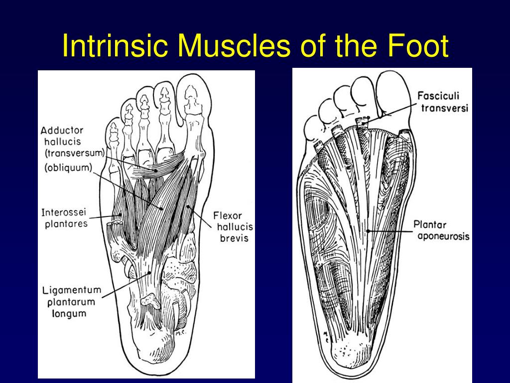
Meanwhile, the task-specific coordination of the foot muscles during other modes of locomotion indicates a high-level of specificity in their function and control. The sequential peak activity of MTP flexors followed by MTP extensors suggests that their biomechanical contributions may be largely separable from each other, and from other extrinsic foot muscles during walking. The intrinsic muscles are much smaller than the extrinsic. Another set of muscles, the intrinsic muscles of the foot, have both proximal and distal attachments within the foot’s bony architecture, from calcaneus to distal phalanx. For instance, extensor hallucis brevis and extensor digitorum brevis muscle activation peaks decoupled during sideways gait.Ĭonclusions. The muscles with proximal attachments at points outside the foot are referred to as extrinsic muscles of the foot. We found that foot muscles that activated synchronously during forward walking tended to dissociate during other locomotor tasks. The period around stance to swing transition could be roughly characterized by sequential peak muscle activity from the ankle plantarflexors, MTP flexors, MTP extensors, then ankle dorsiflexors. We found that peak MTP flexor activity occurred significantly before peak MTP extensor activity during walking (P < 0.001). They are mainly responsible for actions such as eversion, inversion, plantarflexion and dorsiflexion of the. The extrinsic muscles arise from the anterior, posterior and lateral compartments of the leg. We computed stride-averaged EMG envelopes and used the timing of peak muscle activity to assess synchronous vs. The muscles acting on the foot The muscles acting on the foot can be divided into two distinct groups extrinsic and intrinsic muscles. We analyzed surface electromyographic (EMG) recordings of intrinsic and extrinsic foot muscles for healthy individuals during level treadmill walking, and also during sideways and tiptoe gaits. We investigated the coordination of MTP flexors and extensors with respect to each other, and to other ankle-foot muscles. However, based on prior recordings it is unclear if muscles that contribute to flexion/extension of the metatarsophalangeal (MTP) joints are activated synchronously to modulate joint impedance, or sequentially to perform distinct biomechanical functions. The human foot undergoes complex deformations during walking due to passive tissues and active muscles.

This research was performed in collaboration with researchers at the Santa Lucia Foundation in Rome, Italy.

Therefore, the purpose of these studies were to examine the roles of these muscles when an external perturbation was employed in the form of varus, neutral, and valgus-wedged shoes. Excessive pronation may impose stress on the extrinsic muscles of the foot leading to injury. Posted by Karl Zelik on Monday, Novemin News.Įxcited to share that our research article entitled “Coordination of intrinsic and extrinsic foot muscles during walking” has been accepted for publication in the European Journal of Applied Physiology. Running overuse injuries are typically associated with excessive pronation of the foot during stance. Accepted for Publication: Coordination of intrinsic and extrinsic foot muscles during walking


 0 kommentar(er)
